Color Blindness
Also known as Color Vision Deficiency
Medical Disclaimer: Information on this page is for educational purposes only and is not a substitute for professional medical advice, diagnosis, or treatment.
See our Terms and Telemedicine Consent for details.


Overview
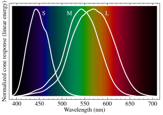
Color blindness —often called color vision deficient (CVD)—is a reduced ability (not usually a total loss) to tell certain colors apart. It happens when one or more of the three cone photopigments in the retina are missing, weakened, or malfunctioning. Red-green problems (protan and deutan types) account for about 95 % of congenital cases, while blue-yellow (tritan) defects and the very rare complete absence of color (achromatopsia) make up the rest.1
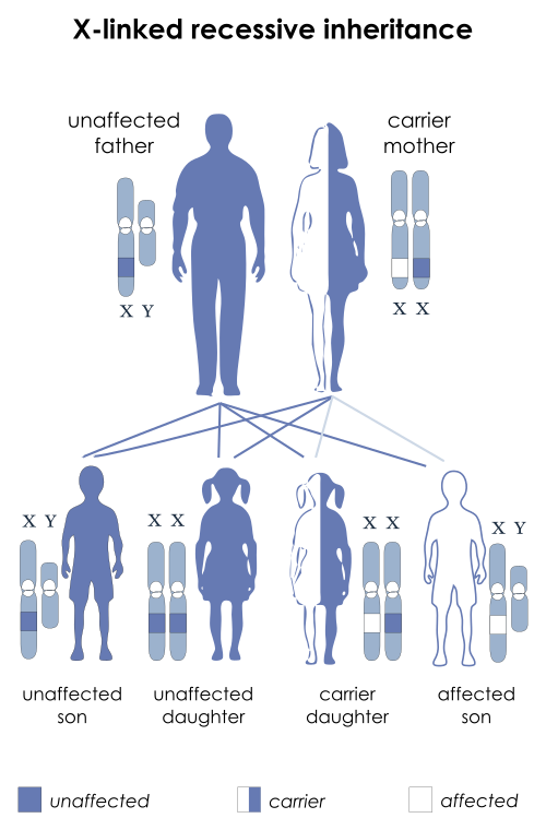
CVD is one of the most common inherited eye conditions—affecting roughly 1 in 12 men and 1 in 200 women of Northern-European ancestry2—because the red- and green-sensitive opsin genes sit on the X-chromosome. While most cases are present from birth, color discrimination can also be lost later in life through retinal disease, optic-nerve damage, medications, or head trauma, so sudden changes in color perception warrant prompt evaluation.3
Symptoms
CVD symptoms vary from subtle to obvious and are influenced by lighting and color saturation:
- Difficulty naming or matching colors—especially reds, greens, browns, and oranges in red-green defects.
- Colors look dull or washed out, with reduced contrast between hues that others see as distinct.4
- Childhood clues: learning color words more slowly, picking the “wrong” crayon, or trouble with color-coded classroom materials.5
- Acquired CVD—caused by eye disease or medicines—may present as a sudden or gradual fading of color intensity, often affecting one eye more than the other.
Visual sharpness is typically normal, so many people are unaware of their deficiency until they take a screening test for school, driver’s license, or an occupation that requires accurate color vision.
Causes and Risk Factors
Congenital CVD results from gene changes that affect cone opsins on the X-chromosome (OPN1LW/OPN1MW) or, less commonly, on chromosome 7 (OPN1SW). It follows an X-linked recessive inheritance pattern—males are far more likely to be affected, while females are usually carriers.6
Acquired or secondary CVD can be triggered by:
- Retinal disorders (e.g., macular degeneration, diabetic retinopathy)
- Optic-nerve diseases (e.g., glaucoma, optic neuritis)
- Medications such as digoxin, hydroxychloroquine, ethambutol, or sildenafil
- Environmental toxins, traumatic brain injury, or stroke
Key risk markers therefore include male sex, family history, Northern-European ancestry, and any co-existing retinal or neurologic disease.2
Color Vision Deficiency Risk Estimator
Select your details to estimate risk factors.
Diagnosis
A comprehensive color-vision evaluation involves:
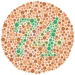
- Ishihara pseudo-isochromatic plates—quickly detect red-green deficiencies and are widely used for screening.7
- HRR plates, Farnsworth D-15, or Farnsworth-Munsell 100-hue tests—classify type and severity, and identify blue-yellow defects.
- Anomaloscope—the gold standard for quantifying red-green anomalies, essential for certain occupational clearances.
- Electroretinography or genetics—occasionally ordered to differentiate congenital from acquired CVD or to confirm rare variants.
Because acquired color loss can signal sight-threatening retinal or optic-nerve disease, any new change should prompt a full dilated exam and ocular imaging.8
Treatment and Management
No cure exists for most inherited forms, but several strategies can improve daily function:
- Adaptive tools—high-contrast labels, textured cues, smartphone apps, and accessible design choices at school and work.
- Special spectral-filter glasses or contact lenses can enhance contrast for some people with mild red-green defects, though they do not restore normal color vision.10
- Underlying-disease management—treat glaucoma, diabetic retinopathy, or medication toxicity to prevent progressive acquired CVD.9
Research shows that notch-filter lenses can improve certain color-naming tasks in red-green CVD, but benefits vary and depth-perception trade-offs exist.11
Living with Color Vision Deficiency and Prevention
Most people with CVD live full lives, but early awareness helps avoid avoidable frustrations:
- Test children by age 4 so teachers can adapt classroom materials and reassure families.13
- Use contrast-friendly design on maps, graphs, and user interfaces—Kerbside practice sheets feature color-safe palettes.
- Protect retinal health: control diabetes, wear UV-blocking eyewear, and review drug side-effects.1
- Occupational guidance: aviation, maritime, and certain technical jobs have color-vision standards—early counseling prevents career setbacks.
Latest Research & Developments
- Gene-replacement therapy using adeno-associated viral vectors restored red-green discrimination in primate models and is moving toward human trials.12
- Wave-guided contact lenses embed micro-filters to augment color contrast without altering overall luminance.
- Machine-vision smartphone apps now translate confusing color codes (e.g., resistor bands, transit maps) into audible cues in real time.
- A randomized crossover study funded by the NEI found that notch-filter spectacles improved color-sorting speed by 15 % in anomalous trichromats.11
Recent Peer-Reviewed Research
How the orientation of the color gamut of natural scenes influences color discrimination in red-green dichromacy.
Marques DN, Nascimento SMC
Empirical tests of the effectiveness of EnChroma multi-notch filters for enhancing color vision in deuteranomaly.
Somers LP, Franklin A, Bosten JM
New pathogenic variants of ALMS1 gene in two Chinese families with Alström Syndrome.
Cheng WY, Ma MJ, Yuan SQ, et al.
Next Steps
Who to see: If you—or your child—struggle with color-related tasks, schedule a comprehensive eye exam with an ophthalmologist experienced in pediatric vision or retinal genetics. They can confirm the diagnosis, rule out treatable causes, and discuss assistive options.3
How to schedule: Many clinics allow self-referral, but your primary-care doctor or optometrist can also generate a referral. Wait times vary; genetically focused practices may book out several weeks, so plan ahead for school or job deadlines.
Need a second opinion or help finding the right subspecialist? You can connect directly with board-certified experts through Kerbside, upload your test results, and receive personalized guidance without leaving home.
Trusted Specialists
Board-certified providers specializing in Color Blindness.


